2 min read

Dr. Ulrike Wegst is a co-author in an article in Advanced Functional Materials titled “X-Ray Tomoscopy Reveals the Dynamics of Ice Templating” published Aug. 21, 2023.
Significance
Biomaterials play an important role for injury repair. For example, when a peripheral nerve has been severed, tubular implants with a honeycomb-like aligned porosity can be used to bridge the gap. These scaffolds provide cells with structural support and guide their directional growth to help the nerve ends reconnect. Freeze casting can be used to fabricate scaffolds, for example, made from chitosan. However, the dynamics of microstructure formation in freeze casting continue to be unknown.
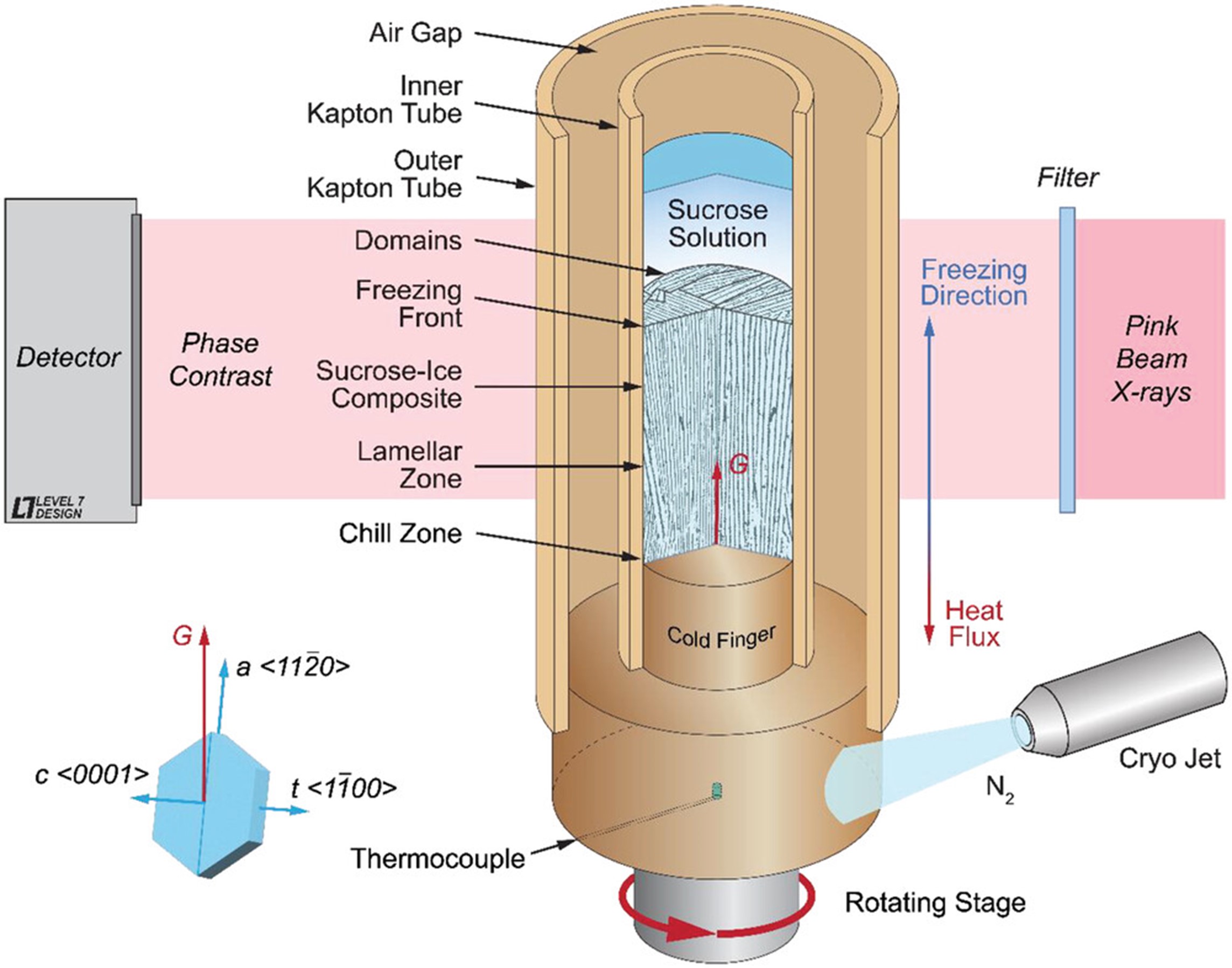
X-ray tomoscopy allowed a real-time visualization of the dynamics of microstructure formation during ice-templating, which forms the basis of the freeze casting process and occur when aqueous solutions or slurries are directionally solidified into cellular solids. Using water-sucrose as a binary model system, the anisotropic, partially faceted crystal growth over sample lengths that include the liquid phase, the mushy zone, and the fully solidified region, in which the solute phase is vitrified, was quntified.
This paper provides evidence of the proposed mechanisms for the evolution of structures based on insights gained through phase field modeling, which was published earlier this year.
Abstract
Little experimentally explored and understood are the complex dynamics of microstructure formation by ice-templating when aqueous solutions or slurries are directionally solidified (freeze cast) into cellular solids. With synchrotron-based, time-resolved X-ray tomoscopy it is possible to study in situ under well-defined conditions the anisotropic, partially faceted growth of ice crystals in aqueous systems.
Obtaining one full tomogram per second for ≈270 s with a spatial resolution of 6 µm, it is possible to capture with minimal X-ray absorption, the freezing front in a 3% weight/volume (w/v) sucrose-in-water solution, which typically progresses at 5–30 µm s−1 for applied cooling rates of . These time and length scales render X-ray tomoscopy ideally suited to quantify in 3D ice crystal growth and templating phenomena that determine the performance-defining hierarchical architecture of freeze-cast materials: a complex pore morphology and “ridges”, “jellyfish cap”, and “tentacle”-like secondary features, which decorate the cell walls.


